
Cover for I Research : Journal of Pharmacology
Open AccessISSN: XXXX-XXXX

I Research : Journal of Pharmacology

The I Research : Journal of Pharmacology provides a resource content about Pharmacology and Toxicology field especially about the pre-clinical studies, Clinical Investigations, Pharmacovigilance etc. This journal aims to publish all the recent research and reviews articles related to pharmacology and Toxicology like Pre-clinical and Clinical studies, PKPD Modelling, Bioavailability studies in animal models, Toxicological studies of synthetic and herbal drugs etc.. It is the international journal of published Bi-Annual by I Research Academia. Authors should consult the latest instructions to authors before preparing their manuscripts. All contributions must be in English and should be submitted online only in a single word file. All contributions must be in English and should be submitted online only in a single word file.

Journal Policy

Current Issue: I Research: Journal of Pharmacology, July - Dec 2026

Volume 2, Number 2 (2026)
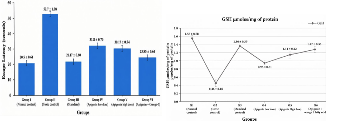
Image for EVALUATION OF THE NEUROPROTECTIVE EFFECT OF APIGENIN AND OMEGA-3 FATTY ACIDS ON CHOLINERGIC DYSFUNCTION AND OXIDATIVE STRESS IN SCOPOLAMINE INDUCED ALZHEIMER’S DISEASE IN RATS
Open AccessResearch Article

EVALUATION OF THE NEUROPROTECTIVE EFFECT OF APIGENIN AND OMEGA-3 FATTY ACIDS ON CHOLINERGIC DYSFUNCTION AND OXIDATIVE STRESS IN SCOPOLAMINE INDUCED ALZHEIMER’S DISEASE IN RATS

By Thiruselvam V, Marihrishnaa K.
Background: Alzheimer's disease (AD) is a progressive neurodegenerative disorder marked by cholinergic dysfunction, oxidative stress, and hippocampal neuronal loss. Scopolamine, a muscarinic receptor antagonist, is widely used to induce reversible cognitive impairment in rodents that reproduces key biochemical and behavioural hallmarks of AD. Apigenin, a naturally occurring flavonoid, and omega-3 fatty acids have each been reported to possess antioxidant and neuroprotective properties, but their combined effect against scopolamine-induced neurotoxicity has not been well characterised. Objective: To evaluate the neuroprotective effect of apigenin, alone and in combination with omega-3 fatty acid, on cholinergic function and oxidative stress in a scopolamine-induced model of cognitive impairment in rats. Methods: Adult Wistar albino rats were divided into six groups (n = 3/group): normal control, scopolamine (1 mg/kg, i.p., days 9–17) toxic control, donepezil (10 mg/kg, p.o.) standard control, apigenin low dose (25 mg/kg), apigenin high dose (50 mg/kg), and a combined apigenin (25 mg/kg) plus omega-3 fatty acid (100 mg/kg) group, all treated orally for 17 days alongside scopolamine. Spatial learning and memory were assessed by the Morris water maze (escape latency), and locomotor activity by digital actophotometer, on days 0, 9, 14, and 17. On day 17, brain acetylcholinesterase (AChE) activity and the oxidative stress markers malondialdehyde (MDA), reduced glutathione (GSH), superoxide dismutase (SOD), and catalase (CAT) were estimated, and hippocampal sections were examined histopathologically (H&E staining). Results: Scopolamine produced significant cognitive and locomotor impairment, elevated AChE activity and MDA levels, depleted GSH, SOD, and CAT, and caused marked hippocampal degeneration compared with the normal control group (p < 0.001). Apigenin treatment attenuated these changes in a dose-dependent manner, and the combined apigenin–omega-3 fatty acid group showed the greatest improvement across behavioural, biochemical, and histopathological parameters, approaching values recorded in the standard (donepezil) group. Conclusion: Apigenin exerts a dose-dependent neuroprotective effect against scopolamine-induced cognitive dysfunction, and its combination with omega-3 fatty acid produces a more pronounced protective response, indicating a complementary antioxidant and cholinergic-restorative action that supports further investigation of this combination as a candidate neuroprotective strategy.
Image for IMMUNOMODULATORY ACTIVITY OF PROPYL GALLATE AGAINST CYCLOPHOSPHAMIDE INDUCED IMMUNOSUPPRESSION IN RAT.
Open AccessResearch Article

IMMUNOMODULATORY ACTIVITY OF PROPYL GALLATE AGAINST CYCLOPHOSPHAMIDE INDUCED IMMUNOSUPPRESSION IN RAT.

By Mukeshkanna R, Dr.N.Chidambaranathan
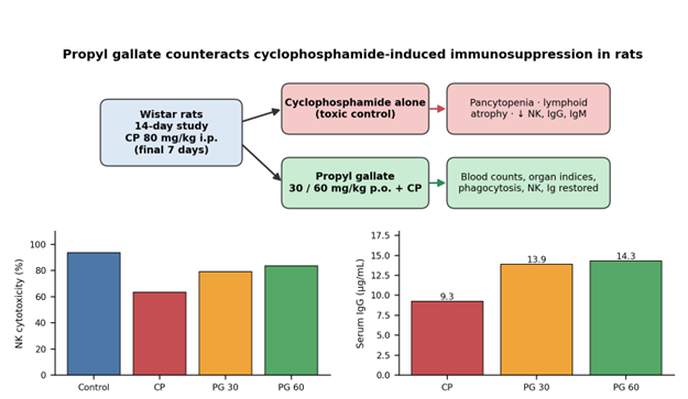
Background: Cyclophosphamide (CP) is a cytotoxic drug with immunosuppressive properties that is used extensively; its clinical value is limited by bone-marrow suppression and damage to lymphoid organs. Propyl gallate (PG), the n-propyl ester of gallic acid, is a phenolic antioxidant reported to have anti-inflammatory and cytoprotective properties, although no report has addressed its influence on CP-induced immune injury. Methods: Wistar rats were divided into five groups: normal control, CP control, levamisole (2.5 mg/kg) + CP, PG 30 mg/kg + CP and PG 60 mg/kg + CP. Test agents were administered for 14 days, with CP (80 mg/kg, i.p.) injected over the last 7 days. End-points comprised blood counts, spleen and thymus indices, carbon-clearance phagocytic index, neutrophil adhesion, splenic natural killer (NK) cell cytotoxicity, serum IgG and IgM (ELISA), and haematoxylin–eosin histology of the spleen and thymus. Results: CP reduced WBC, RBC and platelet counts by 50.5%, 37.0% and 27.0% respectively, approximately halved the spleen and thymus indices, and impaired phagocytosis, NK-cell cytotoxicity and immunoglobulin levels. Compared with CP alone, PG at 60 mg/kg raised WBC (+63%), RBC (+23%) and platelets (+20%), improved the spleen and thymus indices, brought phagocytic capacity back to normal, recovered NK-cell cytotoxicity (83.7% versus 63.4%) and elevated IgG and IgM by 55% and 67%. The 30 mg/kg dose gave smaller and mostly partial responses, with evident improvement in leukocyte counts, immune organ indices, phagocytosis, NK activity and immunoglobulins. Histological examination indicated that PG maintained lymphoid cellularity in the spleen and thymus. Conclusion: In a dose-related manner, PG shielded the haematopoietic, lymphoid and effector-cell compartments from CP, with an effect size close to that of levamisole. These results mark PG as a candidate immunorestorative adjunct worthy of mechanistic, safety and tumour-model investigation.
Image for Comparative Evaluation of Hepatoprotective  Activity of Apigenin and Alpha-Tocopherol Against Methotrexate-Induced Hepatotoxicity in Rats
Open AccessResearch Article

Comparative Evaluation of Hepatoprotective Activity of Apigenin and Alpha-Tocopherol Against Methotrexate-Induced Hepatotoxicity in Rats

By Vignesh .V, Dr.N.Chidambaranathan
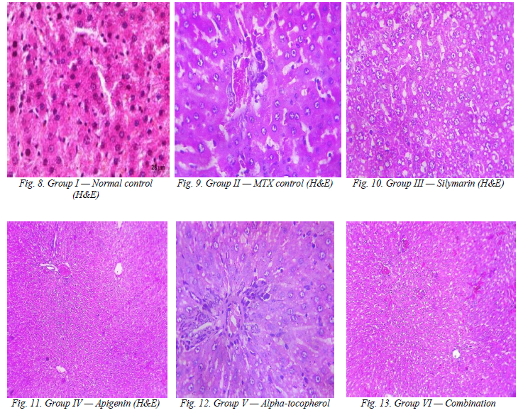
Background: Methotrexate (MTX), a folate-antagonist antimetabolite widely used in the management of malignancies, rheumatoid arthritis and psoriasis, has clinical utility that is frequently limited by dose-dependent hepatotoxicity driven largely by oxidative stress. Objective: This study was designed to comparatively evaluate the hepatoprotective potential of apigenin and alpha-tocopherol, individually and in combination, against MTX-induced hepatic injury in rats, with silymarin employed as the standard reference hepatoprotective agent. Methods: Twenty-four Wistar albino rats were allocated into six groups of four animals each: normal control; MTX control (20 mg/kg, i.p., single dose on day 7); silymarin (50 mg/kg/day, p.o.) plus MTX; apigenin (50 mg/kg/day, p.o.) plus MTX; alpha-tocopherol (100 mg/kg/day, p.o.) plus MTX; and a combination of apigenin and alpha-tocopherol plus MTX. Oral treatments were administered daily for fourteen days. Hepatic superoxide dismutase (SOD), catalase (CAT), reduced glutathione (GSH) and malondialdehyde (MDA), together with serum alanine aminotransferase (ALT), aspartate aminotransferase (AST) and alkaline phosphatase (ALP), were estimated, and liver sections were examined histologically. Results: MTX administration produced a marked depletion of hepatic SOD, CAT and GSH and a significant elevation of MDA, ALT, AST and ALP relative to the normal control (p<0.001), accompanied by hepatocellular degeneration, cytoplasmic vacuolation and pyknotic nuclei on histopathology. Pre-treatment with apigenin, alpha-tocopherol and, most markedly, their combination significantly attenuated these oxidative and biochemical derangements and preserved hepatic architecture (p<0.001 versus the MTX control), with the combined regimen producing the most consistent protection among the test groups and approaching the effect of silymarin. Conclusion: Apigenin and alpha-tocopherol confer hepatoprotection against methotrexate-induced liver injury through complementary antioxidant mechanisms, and their combined use may represent a rational adjunct strategy meriting further pharmacological and clinical investigation.
Image for EVALUATION OF CARDIPROTECTIVE ACTIVITY OF RUTIN PHYTOSOME AGAINST DOXURUBICIN INDUCED CARDIOTOXICITY BY USING ANIMAL MODELS
Open AccessResearch Article

EVALUATION OF CARDIPROTECTIVE ACTIVITY OF RUTIN PHYTOSOME AGAINST DOXURUBICIN INDUCED CARDIOTOXICITY BY USING ANIMAL MODELS

By Karthikeyan M, Jegan N
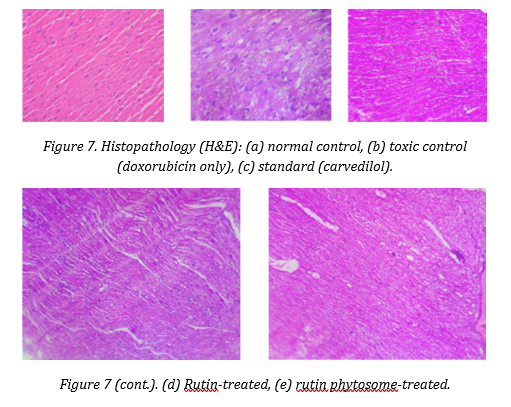
Background: Doxorubicin remains a cornerstone anticancer agent, but its clinical utility is limited by dose-dependent cardiotoxicity mediated largely through oxidative stress and mitochondrial injury. Rutin, a flavonoid glycoside with well-documented antioxidant activity, is constrained therapeutically by poor aqueous solubility and limited membrane permeability. Complexation with phosphatidylcholine to form a phytosome was investigated as a strategy to overcome these limitations and enhance cardioprotective efficacy. Methods: Rutin phytosome was prepared by the thin-film hydration/solvent-evaporation technique at three rutin-to-phosphatidylcholine ratios (1:0.5, 1:1, 1:1.5) and characterised by entrapment efficiency, Fourier-transform infrared (FTIR) spectroscopy, scanning electron microscopy (SEM), and in vitro dialysis-membrane drug release. The optimised formulation was evaluated in vivo in a five-group Wistar rat model of doxorubicin-induced cardiotoxicity (normal control; toxic control, doxorubicin 20 mg/kg i.p.; standard, carvedilol 30 mg/kg; test drug, rutin 50 mg/kg; test formulation, rutin phytosome 50 mg/kg), with cardiac biomarkers (CK-MB, LDH, troponin-I), myocardial antioxidant parameters (SOD, CAT, MDA), and histopathology as endpoints. Results: The 1:1 formulation (F2) gave the highest entrapment efficiency (94.2%). FTIR confirmed retention of rutin's characteristic bands with shifts indicative of hydrogen bonding, and SEM showed irregular, rough-surfaced, predominantly spherical particles. In vitro release was sustained, reaching 83.1% cumulative release at 6 hours. In vivo, doxorubicin produced marked elevation of CK-MB, LDH and troponin-I and depletion of SOD and CAT with elevated MDA, confirming cardiotoxicity. Pretreatment with rutin phytosome produced a greater reduction in all three cardiac biomarkers than plain rutin (CK-MB 34.14 ± 0.49 vs 38.68 ± 0.62 U/L; LDH 253.2 ± 3.83 vs 291.4 ± 3.97 IU/L; troponin-I 0.79 ± 0.037 vs 2.08 ± 0.020 ng/mL) and better restoration of antioxidant status, findings corroborated by graded histopathological recovery of myocardial architecture. Conclusion: Phospholipid complexation improved the physicochemical and biopharmaceutical profile of rutin and conferred superior protection against doxorubicin-induced cardiotoxicity relative to free rutin, supporting further pharmacokinetic and clinical translational evaluation of rutin phytosome as an adjuvant cardioprotective agent.
Image for Green Synthesis of Caulerpa chemnitzia–Derived Silver Nanoparticles for Breast Cancer Therapy: Current Advances, Mechanisms, and Future Perspectives
Open AccessReview Article

Green Synthesis of Caulerpa chemnitzia–Derived Silver Nanoparticles for Breast Cancer Therapy: Current Advances, Mechanisms, and Future Perspectives

By S.Gopi, N. Jegan
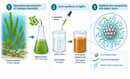
Abstract Breast cancer remains the most frequently diagnosed malignancy among women worldwide and a leading cause of cancer-related death, prompting sustained interest in therapeutic strategies that combine efficacy with reduced systemic toxicity. Green nanotechnology, in which biological extracts replace hazardous chemical reagents as reducing and capping agents during nanoparticle fabrication, has emerged as an environmentally benign route to biologically active nanomaterials. Marine macroalgae of the genus Caulerpa are of particular interest because their tissues are naturally rich in polyphenols, sulfated polysaccharides, pigments, and distinctive alkaloids such as caulerpin, all of which can serve simultaneously as reducing and stabilizing agents during the biosynthesis of silver nanoparticles (AgNPs). This review synthesizes the published evidence on green synthesis of AgNPs from Caulerpa species, including Caulerpa chemnitzia, C. racemosa, C. serrulata, C. scalpelliformis, and C. lentillifera, with an emphasis on synthesis optimization, physicochemical characterization, and the cellular mechanisms by which such nanoparticles exert cytotoxic and pro-apoptotic effects on breast cancer cell lines, principally MCF-7. Reactive-oxygen-species generation, mitochondrial membrane depolarization, caspase-3/9 activation, and modulation of Bax/Bcl-2 expression recur as the dominant mechanistic themes across the literature reviewed. The review further situates Caulerpa-derived AgNPs within the broader landscape of algae- and plant-mediated nanoparticle synthesis, addresses toxicological and biocompatibility considerations, and outlines current gaps — most notably the scarcity of dedicated breast-cancer bioassays for C. chemnitzia-derived AgNPs specifically — together with priorities for future in vivo and translational research.
Image for Evaluation of the Antidiabetic and Antioxidant activities of ochthochloa compressa in STZ-induced diabetic rats.
Open AccessResearch Article

Evaluation of the Antidiabetic and Antioxidant activities of ochthochloa compressa in STZ-induced diabetic rats.

By Bharathi kannan.M, Marihrisnaa.K

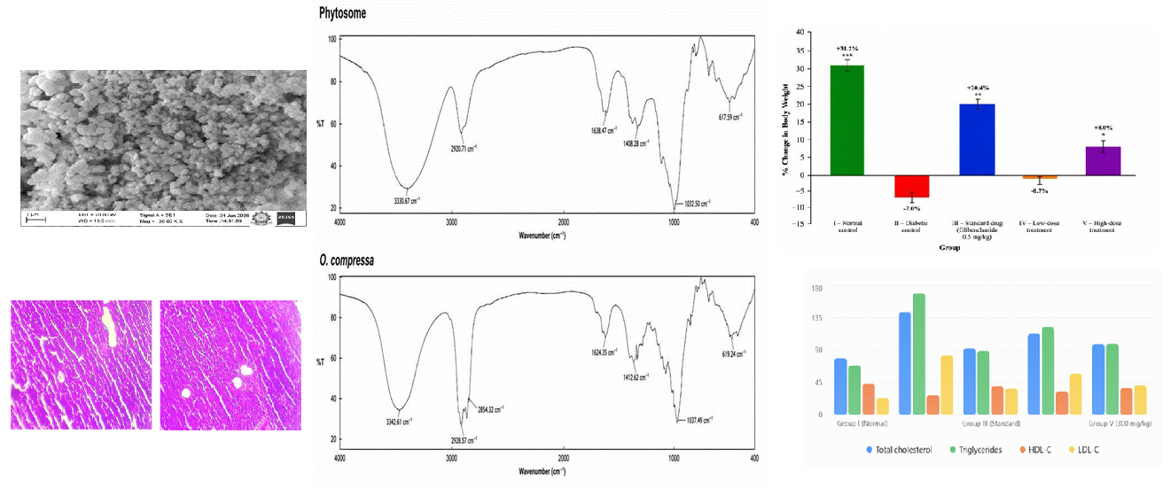
Ochthochloa compressa (Forssk.) Hilu is a medicinal grass of the Poaceae family reported to possess antidiabetic and antioxidant phytoconstituents, but its poor aqueous solubility limits the oral bioavailability of the unformulated extract. This study aimed to formulate a phospholipid-complexed phytosome of O. compressa extract and to evaluate its antidiabetic and antioxidant activity in streptozotocin (STZ)-induced diabetic rats. The phytosome was prepared by the thin-film hydration method using soy phosphatidylcholine and optimised across four extract-to-phospholipid ratios. The optimised formulation (1:3) showed a particle size of 217.2 nm, a polydispersity index of 0.324, a zeta potential of −28.6 mV, and an entrapment efficiency of 89.5%. FTIR confirmed extract–phospholipid interaction, and SEM revealed a nanostructured particulate morphology. In vitro release was sustained, reaching 76.4% at 8 hours. Diabetes was induced in Wistar rats by a single intraperitoneal dose of STZ (60 mg/kg); animals were then treated orally for 15 days with glibenclamide (0.5 mg/kg) or the phytosome (100 or 300 mg/kg). The phytosome produced a dose-dependent reduction in fasting blood glucose, improved body weight and lipid profile, lowered elevated liver and kidney function markers, and restored SOD, catalase, GSH and MDA levels toward normal, with the 300 mg/kg dose approaching the efficacy of glibenclamide. Pancreatic histopathology showed preserved islet architecture in phytosome-treated groups relative to the diabetic control. These findings indicate that phytosome complexation is a promising strategy for improving the antidiabetic and antioxidant performance of O. compressa extract.

News & Announcements

There are no news or announcements at this time.